
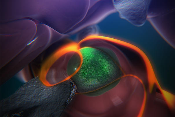

- A still from an animation that shows the steps of how blood stem cells take root in the body of a zebrafish to generate blood. (Credit: Boston Children’s Hospital/Stem Cell Research Program)

A see-through zebrafish and enhanced imaging provide the first direct glimpse of how blood stem cells take root in the body to generate blood. Reporting online in the journal Cell, researchers in Boston Children’s Hospital’s Stem Cell Research Program describe a surprisingly dynamic system that offers several clues for improving bone-marrow transplants in patients with cancer, severe immune deficiencies, and blood disorders, and for helping those transplants “take.”
The steps are detailed in an animation narrated by senior investigator Leonard Zon, director of the Stem Cell Research Program and professor of stem cell and regenerative biology at Harvard Medical School.
“The same process occurs during a bone-marrow transplant as occurs in the body naturally,” says Zon. “Our direct visualization gives us a series of steps to target, and in theory we can look for drugs that affect every step of that process.”
 Credit: Boston Children’s Hospital
“Stem cell and bone marrow transplants are still very much a black box — cells are introduced into a patient and later on we can measure recovery of their blood system, but what happens in between can’t be seen,” says Owen Tamplin, the paper’s co-first author. “Now we have a system where we can actually watch that middle step. “


