Removing tumors accurately during cancer surgery is one of the biggest challenges doctors face.
In breast cancer, for example, up to 35% of patients may still have cancer cells left at the edge of the removed tissue, often leading to repeat surgeries and a higher chance of the cancer coming back.
Traditional imaging methods like ultrasound often fail to show the full extent of the tumor, leaving surgeons to rely mostly on experience.
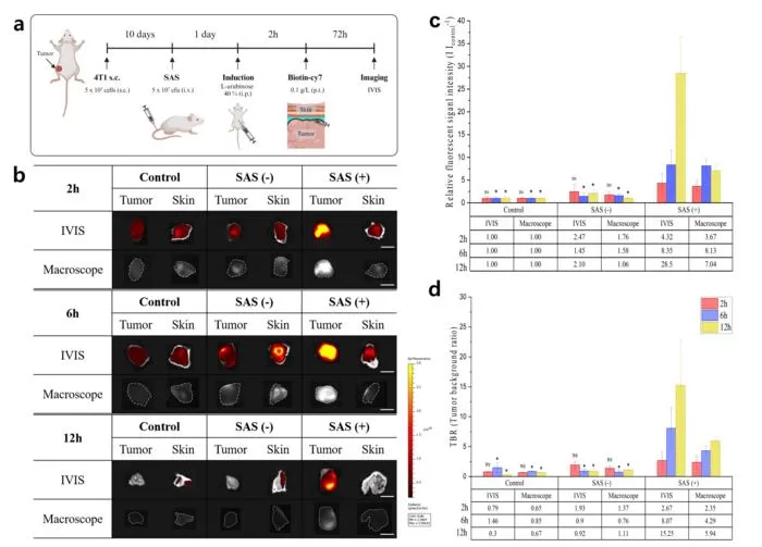
Now, a team of researchers from the Korea Institute of Science and Technology (KIST) and Chungnam National University Hospital has developed a new imaging technology that utilizes engineered bacteria to aid surgeons in clearly visualizing tumors during surgery.
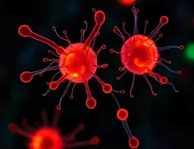
These bacteria are designed to glow only at tumor sites, acting like a neon sign that highlights cancerous tissue in real time.
How It Works
The bacteria activate only in tumor environments and produce a fluorescent signal that lasts for more than 72 hours. This signal is strong enough to be seen with the naked eye—even under normal surgical lighting—and helps doctors identify the exact location and edges of the tumor.
Broad Use Across Cancer Types
Unlike older contrast agents that must be customized for each cancer type, this new system works by detecting common tumor traits like low oxygen levels and immune system evasion. It’s also five times brighter than traditional agents and works in the near-infrared range, making it compatible with existing surgical tools and imaging systems.
Future Possibilities
The team hopes to turn this platform into a complete cancer treatment system. The bacteria could eventually be used to deliver cancer drugs directly to tumors, combining diagnosis, surgery, and therapy in one approach.
Dr. SeungBeum Suh from KIST said, “This study shows a new way to find tumors using bacteria that glow. It could become a new standard for precise cancer surgery.”
This breakthrough could make cancer surgeries safer, more accurate, and less likely to require follow-up procedures

