
Osaka, Japan (RSN)– Autoimmune diseases occur when a person’s antibodies mistakenly recognize their body’s own cells as foreign invaders and attack them. One such disease is myasthenia gravis (MG), where the antibodies target neuromuscular-associated proteins.
MG is common in patients with thymoma, a type of tumor of the thymus, but the reasons for this are unclear. Recently, Osaka University researchers used genetic techniques to examine the relationship between these two diseases. Their findings may pave the way for new treatments for this condition.
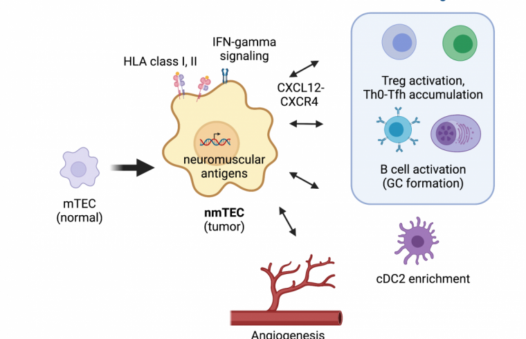
In a recent article published in Nature Communications, the team described how they aimed to identify thymoma-specific changes that occur in cells of the thymus – part of the lymphatic system – to find the connection with MG. A variety of epithelial cells within the thymus work together during various steps of T cell maturation.

T cells are key fighters of the human immune system. One step is mediated by medullary thymic epithelial cells (mTECs), which help prevent the production of self-targeting T cells. Previous studies have shown that immune cells involved in producing antibodies are enriched in MG-thymoma cases, but their role in the effects of MG is not well understood.
At a very basic level, many diseases are ultimately fueled by abnormal gene expression patterns,” says lead author Yoshiaki Yasumizu. “We believed that examining the levels of certain RNA molecules in MG-thymoma samples would help us find specific genes directly involved in this disease.”
Using a method called RNA-sequencing, the team evaluated over 100 thymoma samples and looked for genes that differed in expression levels between cases with and without MG. These experiments helped them identify neuromuscular molecules that were present in higher amounts in MG-thymoma samples.
“Further work showed us that these neuromuscular molecules are expressed in a subpopulation of mTECs, which led us to name them neuromuscular mTECs (nmTECs),” explains Hideki Mochizuki, senior author of the study.
Molecular characterization of the nmTECs suggested that they are capable of a process called antigen presentation, where a molecule is placed at the cell surface to induce an immune response against it. The nmTECs present the neuromuscular molecules, leading to the production of antibodies against them.
“We also observed abnormal follicular structures and T/B cell behaviors, all of which help produce a microenvironment that can support autoimmune outcomes,” says Yasumizu.
By staining certain proteins using a technique called immunohistochemistry, the team was able to confirm the presence and enrichment of nmTECs in MG-thymoma samples. Overall, this study provides strong evidence that nmTECs are significantly involved in MG through the high expression of neuromuscular molecules. This new knowledge may be the key to the development of novel therapeutic methods for this disease combination.


