
In a recent study published in Electrophoresis, University of Malaya’s researchers found significant correlation between increased levels of serum amyloid A (SAA) in patients with three different bone sarcomas: pleomorphic sarcoma (PS), osteosarcoma (OS) and chondrosarcoma (CS) and the different degrees of tumor malignancy in PS, OS and CS.

Credit : Electrophoresis (2016) 37 (17-18): 2328-2337
Sarcomas are rare cancers originating from bone and soft tissue (skeletal system) which often have a predilection for a specific age group. Both pleomorphic sarcoma (PS) and osteosarcoma (OS) are aggressive malignant and highly metastatic tumors while chondrosarcoma (CS) has low metastatic rate and somewhat less aggressive in comparison to PS and OS. PS and CS are usually reported in adult cancer patients but OS affects mainly adolescents and children. Although these sarcomas are very similar in presentation, only OS has been well studied.
Serum amyloid A (SAA) is an apoliproprotein which is synthesized in liver and found in low level in sera of healthy people. SAA is one of the major proteins involved in tumorigenesis and elevated levels of SAA may contribute to increase chances for a tumour to progress and metastasize. Several reports have described significant upregulated levels of SAA in sera of cancer patients including OS patients. Thus, the researchers initiated analysis of the abundance of SAA in patients with PS, OS and CS as well as healthy subjects as control groups. The analysis was done utilizing 2DE, Western blotting, ELISA and multiple-reaction monitoring (MRM) analyses.
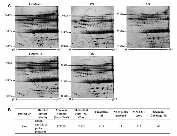
Significant upregulated levels of SAA in patients with PS in comparison to their age-matched control subjects were reported for the first time by the researchers in this study. Initially the results were discovered via combination of 2DE-based proteomics and MALDI TOF/TOF analysis. Later, they were confirmed with results from Western blot and ELISA. Patients with PS recorded the highest levels of SAA followed by patients with OS and CS. The researchers further explored the elevated levels of SAA isoforms in these patients using MRM analysis, where ultraperformance liquid chromatography was coupled with mass spectrometry analysis to monitor and quantify the isoforms. Two of the four SAA isoforms, SAA1 and SAA2, were independently targeted in the MRM analysis and it was found that levels of SAA1 in the sera of PS, OS and CS patients were higher in comparison to SAA2. Previous study had suggested that SAA1 protein acts as an effector for the metastasis-promoting functions of the S100A4 protein, linking the inflammation process with tumour progression.
Furthermore, upregulated levels of SAA, particularly isoform SAA1, were found to be more apparent in patients with PS and OS as opposed to those with CS. This suggested that the contrasting increased levels of SAA in patients with PS, OS, and CS may be related to the different degrees of tumor malignancy. Thus, SAA has a potential to be developed into prognostic biomarker although this requires further validation in clinically representative populations. The levels of SAA should also be clinically investigated for use as a discriminative indicator of treatment efficiency and/or a signal for relapse.


